
Modern microscopes working at the nanotech level need pristine surfaces. Tom Levesque, Technology Business Consulting, and Jezz Leckenby, Talking Science, discuss the use of plasma cleaning for such instruments.
With examination and fabrication nearing the atomic level, the cleanliness of specimen surfaces and the high vacuum electron microscope environments in which these surfaces are studied or processed have never been more critical than they are today. Routine manufacturing at the scale required for nanotechnology demands pristine and controlled surfaces to create the desired structures. Modern electron and ion microscopes are equipped with sophisticated vacuum systems and can provide these conditions, but maintaining cleanliness over time may be more difficult. One of the ways that scientists have been able to achieve these remarkably unadulterated surfaces has been to subject their samples and microscopes to cleaning by various plasma technologies.
Often contamination is derived from hydrocarbon molecules, which even in minute quantities can interact with the electron or ion beam, creating unwanted artefacts in images or data.
The problem of hydrocarbon contamination inside the electron microscope is well documented and has been an issue from the earliest days of electron microscopy. This artefact is the often result of the electron (or ion) beam striking unwanted contaminant molecules and promoting the growth of carbonaceous materials on the surface of the sample. Typically, in a beam-scanning instrument, this contamination layer is in the shape of the rastered pattern on the sample – a rectangle. Further, if the contaminated area is measured in an atomic force microscope (AFM), the build-up of this material has been shown to be quite pronounced.1
Cleaning the specimen before placing it in the scanning electron microscope (SEM) helps, but there is always a small amount of hydrocarbon in the system. Also, the source of these residual hydrocarbons is manifold as they can be left behind from the manufacturing of the tool, part of the vacuum or lubrication system, or derived from the sample or sample handling. Hence, there is the requirement for periodic chamber cleaning.
Previous approaches
Historically, plasma cleaners have been adapted from plasma ashers where the goal was to remove all the organic materials. Commonly used in the semiconductor industry to remove resistive materials composed of polymeric hydrocarbons, these large systems gave rise to smaller, laboratory units that were used to clean samples and parts often destined for examination in vacuum environments such as electron microscopes and other surface analysis tools (see Fig. 1).
Primary plasma cleaning systems
Traditional plasma cleaning systems may be categorised as primary plasma systems. These create gas plasma via an energy source that ionises and dissociates a source gas, resulting in physically and chemically active components. Samples intended for cleaning are placed directly into the gas discharge, on or near the electrode plates of the system and receive full exposure to all the aggressive species of the plasma (i.e. ions, free radicals and by-products).
The original work by Zaluzec2 resulted in a patent and led to commercial products that first used direct plasma to clean samples and parts destined for use in electron microscopes. This has allowed scientists to remove substantial contamination artefacts from their images.
Follow-up work, such as that of Isabel and Fischione,3 continued to demonstrate the value of plasma cleaning to remove problematic hydrocarbons and even to clean contaminated samples if they have already suffered beam-induced carbonaceous polymerisation on their surfaces. Most of these direct cleaners use argon or argon/oxygen mixtures. In these systems, the samples are directly exposed to the energetic ions, and even at low power (10–20W) there is the possibility of surface ablation and damage of sensitive samples such as lacy carbon films.
Plasma cleaners are now common in the electron microscope (EM) suite, with users in many disciplines cleaning both samples and sample holders prior to microscopy. Direct plasma cleaners are available from several manufacturers. These vary both in sophistication and price. However, these instruments do not address the internal surfaces of the microscope. Although the amounts of mobile hydrocarbon contaminants contained by those internal surfaces might be miniscule, the scales at which observations are now being pursued are such that the issue requires an effective resolution if progress is to be maintained.
Any proposed solution that rests upon a requirement for the disassembly and manual cleaning of components is unlikely to be practical and – even if pursued – would entail a level of manual intervention that would potentially place a disproportionate economic burden on any assessment of research costs and benefits.
Downstream systems
In 1999, XEI Scientific patented and introduced a radio frequency (RF) plasma product that used the technique of secondary or downstream plasma cleaning to address the problem of cleaning internal surfaces of vacuum chambers in electron microscopes. A schematic of this downstream plasma process is shown in Figure 2. This type of system produces the active plasma in a remote chamber (called a Plasma Radical Source or PRS) and transfers the active species to the cleaning chamber via gas flow, relying primarily on the chemical activity of the reactive radicals produced by the plasma.
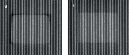
Figure 3: An illustration of the effects of plasma cleaning on a NIST reference sample before (a) and after (b) treatment
Image courtesy of NIST
Experiments with different gases to create the plasma have shown room air to be an excellent source of oxygen to create reactive radicals and crack hydrocarbon molecules efficiently. It has the benefits of being available, free and safe. An illustration of the effects of plasma cleaning on a NIST reference sample is shown in Figure 3, before (a) and after (b) treatment.
This downstream plasma cleaning technique has proven to be extremely useful in electron and ion column instrumentation as the technique can remove unwanted hydrocarbon contamination from the insides of complex instrumentation without disassembly. An example is the Evactron PRSv equipped with a KF40 flange and impedance matching electronics configured for SEM or dual beam FIB use (Figure 4).
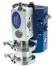
Figure 4: Downstream plasma cleaning system
Image courtesy of XEI Scientific
A further benefit of this technique is that its benign nature has proven safe for common but sensitive materials found inside electron microscopes (such as X-ray detector windows), and has been used for years with no adverse effects observed by a very large number of researchers.
Plasma radical sources are now available in a number of configurations for most makes and models of electron and ion microscopes. Standard PRS units commonly allow for air or oxygen and oxygen/argon mixtures. These units have KF 40 flanges adapted for most SEMs and Dual Beam FIB/SEMs. Further, because they are portable, these systems may be used to clean a number of different electron microscopes in the laboratory.
A high vacuum Conflat flange version is available for surface analysis tools or other high vacuum chambers and provides for the use of hydrogen gas. Versions of the PRS specific for TEMs where a patented hollow cathode is inserted through the sample insertion port are in final design and testing with most major TEM suppliers. This unique design delivers the cleaning capability directly to the hard-to-reach area where beam and sample interact in the TEM (Figure 5).
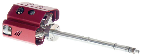
Figure 5: TEM Wand
Image courtesy of XEI Scientific
Downstream plasma cleaning in metrology
Workers at National Institute of Standards and Technology (NIST) in Washington, DC are strong believers in removing all contamination from both samples and chambers. The work of the NIST nanoscale metrology group needs highly accurate scanning electron and helium ion microscopy down to sub 1nm resolution, and this demands contamination-free operation.
Repeatable results require a high degree of cleanliness so that over the few minutes of measurement, the sample does not change noticeably. This also includes the need for consistent secondary electron emission (yield).
Cleaning regimes utilising a liquid nitrogen trap, clean nitrogen gas bleeding, cryo, special pump oil, oil free methods and others had all been deployed at NIST without reaching the standards required by the Institute.
However, the downstream plasma cleaning method that is deployed at NIST has led to the adoption of the technology – using the downstream plasma technique – since it has been assessed as the only current mechanism that will allow the NIST contamination specifications to be met. NIST does not endorse a specific product or brand, but is a leading advocate of the use of plasma-based SEM cleaning.
Implementation and regular use of these methods has made it possible to eliminate the effects of electron beam induced contamination.
The images in Figure 6 were generated by Andras Vladar at NIST.4 They illustrate carbon contamination on a sample in a scanning electron microscope. Images i and iii show the build-up after scanning for 10 minutes at 5kV and 10pA. Image ii shows the effect of using a cryo trap. Contamination is reduced but is still clearly present. Contrast this with the lower pair of images. The last image (iv) shows the result of using downstream plasma cleaning with almost no contamination observed.
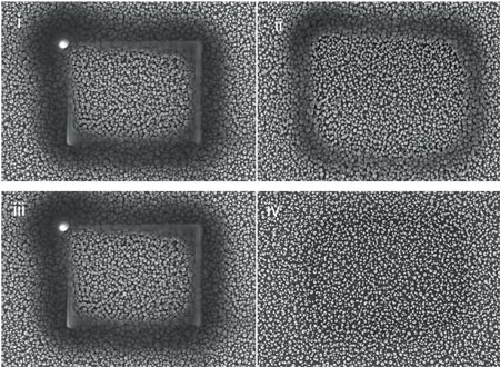
Figure 6: Carbon contamination – examples of cleaning. (i) shows contamination build-up; (ii) contamination reduced with use of a cold trap; (iii) contamination build up; and (iv) almost complete prevention of contamination
Images courtesy of NIST
Other applications
Critical dimension measurements
Comparison imaging to examine the effects of contamination on critical dimension (CD) measurements has shown that these image artefacts can affect dimensional measurements.4
In CD work, modification of dimensions by the SEM imaging process causes a loss of precision in the measurement. Using a very clean Hitachi 6280, the test pattern (Figure 7a) began to show filling-in of the holes after a 20-minute scan. After in situ cleaning of the chamber and the specimen, a repeat of the measurement showed no filling of the holes and a much-reduced scan mark (Figure 7b).
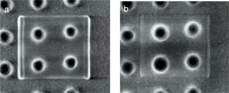
Figure 7: Holes before (a) and after (b) cleaning
Images courtesy of NIST
Analytical microscopy and spectrometry
Horiuchi et al5 have also shown that analytical transmission electron microscopy (TEM) results on polymer brush samples could be accomplished with a system cleaned using downstream plasma. Electron energy loss spectrometry (EELS) in imaging mode could be used for high-resolution carbon mapping.
Nanoscale applications
Having pristine surfaces is an absolute requirement for nanomanipulation and nanofabrication. The work by Mancevski6 has shown that downstream plasma cleaning was essential for successful vapour phase cutting of carbon nanotubes using a nanomanipulator system. Also, electrical measurements made by positioning minute probes on circuits, using nanopositioning systems located inside SEMs and FIBs, require that the probes be free of contamination to make good contacts. In situ cleaning of these devices is a requirement for accurate measurements and plasma cleaners are considered almost an essential accessory for nearly all of these new tools.
In conclusion, downstream plasma cleaning has evolved into a very effective method to get the best possible images and analytical data from sophisticated electron and ion column tools. These systems have proved to be a reliable technique to remove problematic hydrocarbon contamination from samples, holders and microscopes themselves.
This downstream plasma cleaning technology is allowing researchers in various fields to optimise performance from their microscopes and investigate, image, analyse and manipulate materials. XEI alone can account for more than 1,300 installations of its tool on nearly all makes and models of SEM and Dual Beam FIB/SEMs. Today most new high-resolution tools come equipped with some form of downstream plasma cleaning upon delivery. Further, service personnel often carry a portable version of the system when they make service and preventative visits in the field to maximise SEM performance.
References
1. M Amman et al, J Vac Sci Technol B 14(1) (1996) 54–62
2. Simultaneous Specimen and Stage Cleaning Device for Analytical Electron Microscopy, US Patent # 5,510,624, Argonne National Laboratory and the University of Chicago, 1996
3. T C Isabell et al, Micros Microanal 5 (1999) 126–35
4. A Vladar, NIST, personal communication
5. S Horiuchi et al, ACS Nano 3(5) (2009) 1297–1304
6. V Mancevski and P D Rack, Vapor Phase Cutting of Carbon Nanotubes Using a Nanomanipulator Platform, MS&T 2010 Conference & Exhibition, Houston, TX