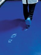
Time-lapse photography has been used to demonstrate the antimicrobial abilities of Dycem contamination control flooring. Dr Gareth Robinson, University of the West of England, Faculty of Health and Life Sciences, explains the procedure and results.
An experiment was designed and carried out with the aim of showing the antimicrobial efficiency of Dycem’s contamination control flooring in comparison with two other commonly used flooring products. For this experiment, Dycem CleanZone polymeric floor covering was used as the first sample; the second comparative sample used was a leading brand of antimicrobial peel-off mat with 30 tabbed sheets; and the third sample was a standard vinyl tile.
A defined volume of bioluminescent reporter bacteria was deposited onto 20mm x 20mm coupons of the Dycem flooring, peel-off mat and vinyl tile. Then by monitoring bacterial bioluminescence, the metabolic inhibition was observed on all three surfaces. The effect occurred more rapidly on Dycem flooring than on the peel-off mat or vinyl. Recovery counts after 3hrs of exposure to the floor surfaces showed a decrease of between 65% and 100% in the number of viable organisms recovered from Dycem flooring compared with the controls.
On vinyl and the peel-off mat, there were slight increases in the number of viable survivors recovered compared with the controls. Dycem flooring inhibited the metabolic activity and reduced the number of viable survivors recovered of Salmonella enterica Serovar Typhimurium DT104 pGLITE. It remains unclear as to whether this effect is attributable to the impregnated antimicrobial or rapid drying of the inoculum on this surface.
The method
Bacterial growth conditions and media: Escherichia coli O157:H7tox-pLITE and Salmonella enterica Serovar Typhimurim DT104 pGLITE were cultured on nutrient agar at 37°C supplemented with 10mg/L ampicillin or 10mg/L gentamicin respectively. Antibiotic selection was used to maintain the plasmid-borne lux genes and the bioluminescent phenotype. Overnight cultures were prepared in 10mL nutrient broth, supplemented with 10mg/L ampicillin/gentamicin as appropriate and incubated at 37°C in an orbital shaker at 200rpm.
Preparation of surface materials: Dycem antimicrobial flooring, the peel-off mat and vinyl tiles were cut into 20 x 20mm coupons. The adhesive surface of the peel-off mat was revealed immediately prior to inoculation. The test surfaces were clean and dry at the time of inoculation. Where recovery counts were performed, the coupons were sterilised using 100% ethanol followed by air-drying and UV irradiation.
Preparation of inoculum: 100µL from an overnight culture was subcultured into 55mL fresh, pre-warmed nutrient broth supplemented with selective antibiotic in a sterile Erlenmeyer flask. The flask was incubated at 37°C in an orbital shaker at 200rpm until an optical density at 600nm (OD600) of 0.4–0.6 was achieved. 50mL of the culture was centrifuged at 4100rpm for 20min at 20°C. The resultant pellet was resuspended in 50mL of fresh nutrient broth or sterile Ringer’s solution as appropriate, or in a tenfold lower volume of nutrient broth for imaging purposes.
Inoculation: The test materials were carefully inoculated with a defined volume of bacterial suspension. The inoculum was placed on the material surface as a single droplet. For imaging experiments, the test coupons were arranged in a square array left open to the air. For recovery count experiments, sterilised coupons were arranged in sterile petri dishes.
Following inoculation the lids were replaced and the plates left undisturbed in a class II microbiological safety cabinet with the fan turned off or on to provide still or moving air around the dish.
Data collection: For imaging experiments, a single 60sec integration was captured every 4min to produce a series of still images over approximately 6.5hrs using an iXonEM+ DU-897BV imaging system (Andor, Belfast, UK). The still images were used to produce the videos appended to the report. Raw data was also analysed to show the decrease in light output over time.
For recovery count experiments, the inoculated coupons were transferred to 50mL Falcon tubes containing 20mL buffered peptone water supplemented with 10mg/L selective antibiotic (recovery diluent) and transferred to an orbital shaker shaking at 200rpm for 20min. The recovery diluent was diluted 1:10 in sterile Ringer’s solution and triplicate recovery plates were produced using a Whitley Automatic Spiral Plater (Don Whitley Scientific, Shipley, UK). Plates were incubated overnight at 37°C followed by recovery counts.
Experimental schedule: The material samples were inoculated with the defined volume of bacteria listed as follows: • Imaging experiment 1. E. coli pLITE suspended in nutrient broth and Ringer’s solution. 20µL deposited volume. •Imaging experiment 2. S. enterica sv. Typhimurium pGLITE suspended in nutrient broth and Ringer’s solution. 50µL deposited volume. •Imaging experiment 3. S. enterica sv. Typhimurium pGLITE suspended in nutrient broth. 50µL deposited volume. •Imaging experiment 4. S. enterica sv. Typhimurium pGLITE suspended in nutrient broth. Inoculum concentrated tenfold. 50µL deposited volume. •Imaging experiment 5. S. enterica sv. Typhimurium pGLITE suspended in nutrient broth. Inoculum concentrated tenfold. 50µL deposited volume. •Recovery count experiment 1. S. enterica sv. Typhimurium pGLITE suspended in Ringer’s solution. 50µL deposited volume. 3hr contact time. Still air. •Recovery count experiment 1. S. enterica sv. Typhimurium pGLITE suspended in Ringer’s solution. 50 µL deposited volume. 3hr contact time. Moving air.
The results: The initial imaging experiment using E. coli O157 pLITE resulted in images with a poor signal-to-noise ratio that were not suitable for conversion to video format. It was decided to utilise S. enterica sv. Typhi-murium pGLITE as it has proven to be a consistently bright reporter strain.
The initial imaging experiment used this organism suspended in nutrient broth and Ringer’s solution to determine the most suitable medium for subsequent imaging work. Raw data taken from the video images are shown in Figs. 1 and 2 for Ringer’s solution and nutrient broth respectively.
In Ringer’s solution, and indeed throughout the experimental run work, there are differences in the initial light output from each floor surface. This is due to the differing coloration of the surfaces. Dycem flooring, being the darkest in colour, absorbs incident light whereas the supplied vinyl flooring was very light in colour, reflecting incident light towards the lens.
The key factor in assessing the data is not the starting light level, but the rate of decline. All three surfaces tested showed a similar initial decline in light output (Fig. 1), but on Dycem flooring light levels were reduced to background within approximately 90min. The peel-off mat showed a very similar initial drop in light, however the rate of decline slowed and a drop to background was not achieved until approximately 4.5hrs. On vinyl flooring the decline in light was initially rapid, slowing at just after one hour and reaching background levels after about 4hrs.
Reduction to background levels took approximately the same time when the inoculum was suspended in nutrient broth (Fig. 2), however light levels remained higher for longer. Light levels on Dycem flooring were reduced to background levels substantially faster than on the peel-off mat or vinyl tile, suggesting that metabolic inhibition occurs much more rapidly on Dycem flooring.
Prior to the current work commencing it had been observed that the physical nature of Dycem flooring encouraged liquid to spread rather than remain as droplets. Following the completion of imaging, all samples had dried out. Video 1 showed that the droplets placed on Dycem flooring spread before the cessation of light emission whereas on the other two surfaces the droplet remained intact. It is not clear whether the rapid metabolic inhibition on Dycem flooring is due to the impregnated antimicrobial, accelerated drying on this surface, or a combination of both.
Three subsequent imaging experiments were conducted using S. enterica sv. Typhimurium pGLITE in nutrient broth alone as this offered higher light levels than suspension in Ringer’s solution. Fig. 3 shows that the pattern of decline in light output is the same as described above. Inhibition of metabolic activity occurs substantially faster on Dycem flooring and on vinyl tile slightly faster than the peel-off mat.
Video 2 showed the same rapid spreading of the inoculum followed by a decline in light in Dycem flooring observed previously.
Inhibition by drying
Figures 4 and 5 represent the data gathered from Video 3 and 4. These were repeats of the previous imaging experiment, however the bacterial inoculum had been concentrated tenfold prior to inoculation to improve further the signal-to-noise ratio. The increased inoculum does not affect the time taken for bioluminescence to decrease to background levels, suggesting that drying is a factor in the metabolic inhibition observed as the deposited volume was unchanged. Both video files clearly show that metabolic inhibition occurs more rapidly on Dycem flooring than on its competitors.
Figs. 6 and 7 show the results of recovery counts when S. enterica was inoculated onto the flooring surfaces and left undisturbed for three hours. In both cases, more bacteria were recovered from the vinyl tile and the peel-off mat than were present in the control.
Since the inoculated samples were covered, there was an element of protection from desiccation not present during the imaging experiment. This may have allowed for limited bacterial growth on the flooring, or growth within the recovery diluent at the end of the contact period. On Dycem flooring, however, bacterial numbers were reduced by more than 65% (Fig. 6) when left in undisturbed air. On these flooring samples, the inoculum had not dried out but it had spread by the end of the experimental period, whereas the inoculum on vinyl tile and the peel-off mat remained as an intact droplet.
Where the class II safety cabinet was operational (Fig. 7), even though the plates were covered, the inoculum had completely dried on the Dycem flooring. In this instance there were no viable survivors recovered from Dycem flooring. On the peel-off mat and on vinyl, numbers of survivors were once again slightly higher than the control.
In conclusion, the experimental work undertaken has shown that metabolic inhibition of S. enterica occurs more rapidly on Dycem flooring than on either vinyl tile or the peel-off mat. Recovery counts have shown that fewer viable bacteria were recoverable from Dycem flooring than from vinyl tile or the peel-off mat.
From the limited nature of the experimental work conducted it is not possible to ascertain whether the inhibitory effect of Dycem flooring is due to the impregnated antimicrobial or the more rapid spreading and drying of liquids on the surface of Dycem.
Contact
Dycem Limited (UK) Ashley Trading Estate Bristol BS29BB UK T +44 117 9559921 .(JavaScript must be enabled to view this email address) www.dycem.com/cr


